In the most delicate and exacting of procedures, world-leading Interventional Neuroradiologist Dr Hal Rice routinely saves lives at the Gold Coast University Hospital (GCUH) – and now with ground-breaking help from 3D printing experts in the Precinct is set to train specialists from around the world.
Dr Rice, together with colleague Dr Laeticia de Villiers, extracts blood clots from inside blocked blood vessels in stroke patients and repairs fragile brain aneurysms that have ruptured or are at high risk of rupturing with catastrophic brain haemorrhage.
The specialist in minimally-invasive endovascular neurosurgical procedures navigates a series of tiny plastic tubes from the femoral artery in the patient’s groin or radial artery in the wrist up into brain blood vessels measuring only two to three millimetres wide, to gently remove blood clots or reconstruct swollen and ruptured blood vessels using innovative devices such as ultra-fine platinum coils and vascular stents, without the need to cut through the skull.
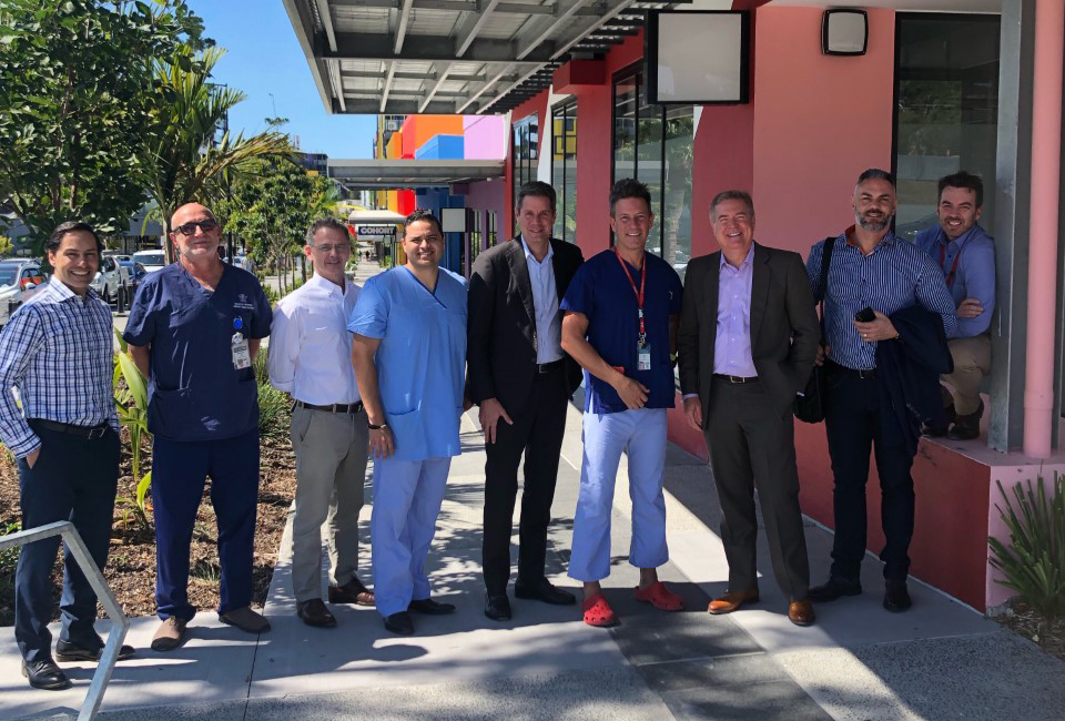
Many of the devices are made by leading global medical company Stryker, with Dr Rice’s world-class standing attracting Stryker’s President of Neurovascular Mark Paul and company executives from the region to visit the Precinct, with a view to using it as their Asia-Pacific base for specialised training.

3D printed models take planning and training to new levels of precision
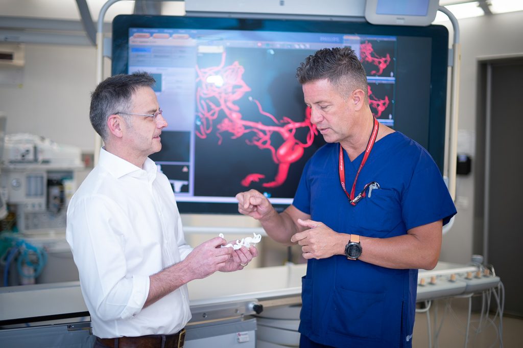
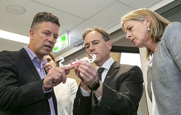
Working with advanced imaging and the specialist digital design skills of Griffith University ADaPT (Advanced Design and Prototyping Technology) experts, they’re 3D printing exact replicas of an individual patient’s aneurysm in situ within the blood vessel so they can better plan life-saving surgeries and train other specialists in this precision specialised medicine.
Conventional training has relied on animal models – the high-tech approach blending virtual simulation with replica printed models will be world-first.
‘The 3D printed models help us to very realistically simulate these complex lifesaving procedures, bringing to life what we see on screen in the operating theatre during an actual treatment,’ says Dr Rice.
‘With a large inventory of precisely printed 3D models we can now rehearse treatment plans and also train specialists in the latest technologies, while using the models to rigorously evaluate new and future products before commencing clinical trials. The models can even be fitted with special pumps to realistically mimic normal pulsating blood flow.’

For Dr Sam Canning, Convenor of Digital and 3D Design at Griffith University, the project tests the limits of design and prototyping technologies.
‘With a project of this complexity we are breaking new ground. We’ve conducted exhaustive tests of combinations of imaging technologies, 3D modelling/imaging software and extensive exploration of 3D printing technologies,”Dr Canning says.
I think it is safe to say that it is recent advances in imaging, software and hardware that have made this entire project possible. Most of this technology (in its current form) did not exist only eighteen months ago.”